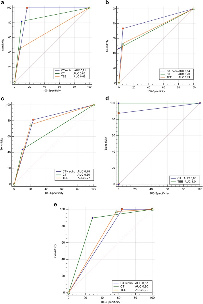
Fig. 6.
Receiver-operating characteristic curves for transesophageal echocardiogram and electrocardiography-gated contrast- enhanced cardiac computed tomography in the diagnosis of abscess/pseudoaneurysm (a), inflammatory infiltration (b), perforations (c), perivalvular leak (d), and vegetations (e). CT, computed tomography; TEE, transesophageal echocardiography; AUC, area under curve

