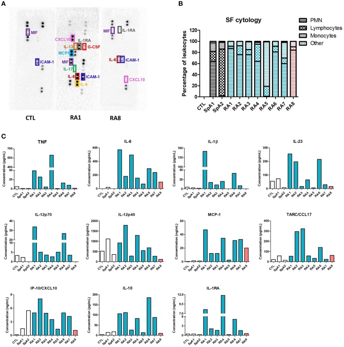
Figure 1.
Evaluation of synovial fluids inflammatory status. SF were obtained from 8 different RA patients, 2 SpA patients and one control SF from a non-rheumatologic joint. RASF are listed depending on their inflammatory status (blue being very pro-inflammatory, orange slightly inflammatory). (A) Pro-inflammatory mediators were identified using an antibody-based membrane array in CTL, RA1, RA8 synovial fluids. Black spots on membrane represent each a cytokine and are semi-quantified in duplicates. Upper and lower rows represent reference spots. (B) SF cytology was determined after routine laboratory analysis. (C) TNF, IL-1β, IL-23, IL-12p70, IL-12p40, MCP-1, CCL17, CXCL10, IL-10, and IL-1RA were quantified in each SF by LegendPlex, MCP-1 was measured by Cytometric Bead Array and IL-6 by ELISA.

