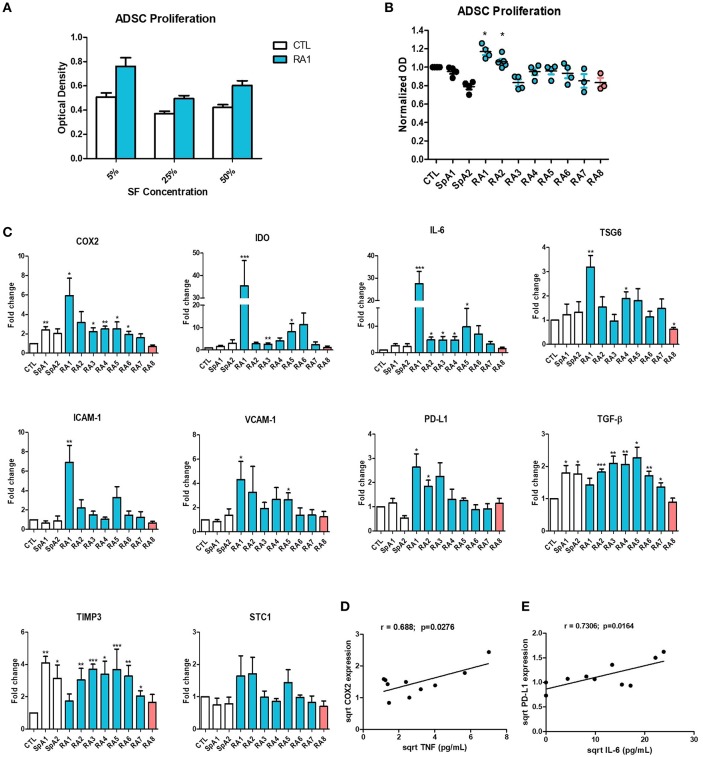
Figure 2.
Differential effect of synovial fluids on ADSC proliferation and gene expression. (A) ADSC were cultured for 48 h in the presence of increasing concentrations of SF Control and RA1 at 5, 25, and 50% (n = 3). (B) For the remaining experiments, ADSC were cultured in the presence of 5% SF for 48 h from either RA, SpA, or control patients (n = 3 − 6). ADSC proliferation was assessed using a colorimetric MTT proliferation assay. (C) ADSC were cultured for 24 h in the presence of SF from either RA, SpA, or control patients. COX2, IDO, IL-6, TSG6, ICAM-1, VCAM-1, PD-L1, TGF-B, TIMP3, and STC-1 gene expression was evaluated by quantitative RT-PCR. RNA levels were normalized to RPS9 (n = 5). Results are represented as mean ± SEM. *p < 0.05; **p < 0.01; ***p < 0.001. (D,E) Correlation (Pearson) between (D) TNF concentrations and COX-2 gene expression and (E) IL-6 concentrations and PD-L1 gene expression. Data are square root-transformed.

