Figure 3.
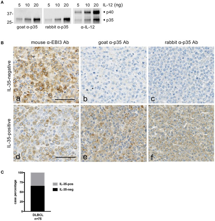
Expression of IL-35 by tumor cells in DLBCL. (A) Specificity of the anti-p35 antibodies used for immunohistochemical studies. Goat or rabbit polyclonal anti-p35 (α-p35) antibodies were tested by western blot using the indicated amount of recombinant IL-12. Anti-IL-12 (α-IL-12) antibody was used as a positive control to detect p35 and p40 subunits. The position of molecular weight standards is indicated on the left (in kDa). (B) Serial sections of DLBCL tissues were stained with anti-EBI3 or anti-p35 antibodies as indicated. Representative cases classified as “IL-35-negative” and “IL-35-positive” are shown. The bar represents 50 μm. (C) Graph indicating the percentage of IL-35-negative and -positive cases among the DLBCL tested by immunohistochemistry.

