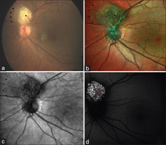
Figure 1.

Color fundus photography of left eye (a) showing creamy white, semi-translucent, well-circumscribed, elevated lesion superior to the optic disc (black arrow). Multicolor imaging (b) highlighted mulberry appearance of the lesion (black arrow) and green shift corresponding to entire extent of tumor mass (arrow heads). In infrared reflectance (c), lesion was hyporeflective with its margins well delineated (arrow heads). Fundus autofluorescence (d) showed typical hyperautofluorescence
