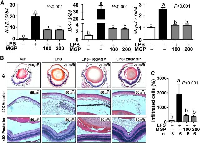
FIGURE 2.
Ocular inflammation and leukocyte infiltration in control and MGP-supplemented C57BL/6 mice. Panel (A) shows proinflammatory gene expression of Il-1β, Il-6, and Mcp-1from enucleated eyeballs. Leukocyte infiltration was visualized by hematoxylin and eosin staining (B). Representative images of the entire eyeball section (top), anterior chamber (middle), and posterior chamber (bottom) are shown. Panel (C) shows the relative leukocyte recruitment (%) into the inflamed eyeball. Values are means ± SEMs (A, C). Values without a common letter differ, P< 0.05; n= 4 for panel (A), and the number of eyes for panel (C) is denoted under the figure. + and − indicate the presence or absence of LPS and/or MGP treatment. IL-1β, interleukin-1β IL-10, interleukin-10; LPS, LPS-injected eyes without supplementation; LPS+100MGP, LPS-injected eyes after 100-mg/kg body weight muscadine grape polyphenol supplementation; LPS+200MGP, LPS-injected eyes after 200-mg/kg body weight muscadine grape polyphenol supplementation; Mcp-1,monocyte chemotactic protein-1; MGP, muscadine grape polyphenol; Veh, vehicle-injected eyes without supplementation.

