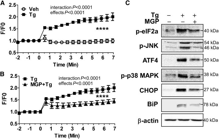
FIGURE 5.
Tg-induced [Ca2+]i? and ER stress markers expression in MGP-treated ARPE-19 cells. Changes are shown in [Ca2+]i?-sensitive florescence concentrations over time in response to vehicle or Tg alone (A) and Tg with MGP treatment (B) in ARPE-19 cells. ER stress-related protein expression of p-eIF2α, ATF4, p-JNK, p-p38 MAPK, BiP, and CHOP by western blot analysis is shown in panel (C). In panels (A) and (B), values are means ± SEMs; n= 5, ****time effect, P< 0.0001; treatment effect, P< 0.0001. In panel (C), β-actin was used as a loading control. + and − indicate the presence or absence of prior treatment of Tg or MGP (100 μg/mL). ARPE-19, human retinal pigmented epithelium; ATF4, activating transcription factor 4; BiP, binding of immunoglobulin protein; CHOP, CCAAT/enhancer-binding protein homologous protein; ER, endoplasmic reticulum; F/F0, ratio of calcium-specific fluorescence; MGP, muscadine grape polyphenol; p-eIF2α, phosphorylated-eukaryotic translation initiation factor 2α p-JNK, phosphorylated c-Jun N-terminal kinase; p-p38 MAPK, phosphorylated-p38 MAPK; Tg, thapsigargin; Veh, vehicle-injected eyes without supplementation; [Ca2+]i?, intracellular calcium.

