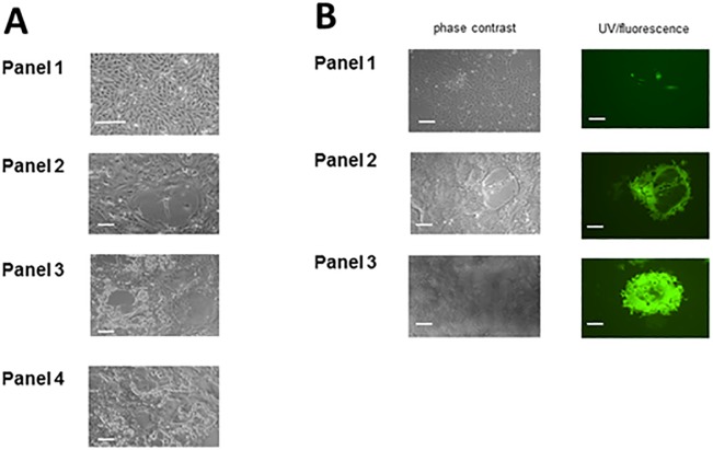
Fig 2. Rescue of the viruses as replicators; cpe and fluorescent plaques.
A: Generation of rMuVG09 by reverse genetics. Panel 1 shows mock infected Vero cells; panel 2 shows the presence of primary foci of rescue at 5 days post transfection; panel 3 shows primary syncytia which were plaque picked and subsequently Vero cells were infected with the aspirated virus stocks. CPE was detected 1–2 dpi. Panel 4 shows plaque picked rMuVG09 grown for 4 low MOI passages on Vero cells. All show characteristic syncytium-formation (scale bar is 50μ). B: Generation of rMuVG09 expressing EGFP—rMuVG09EGFP(3)—by reverse genetics. Panel 1 shows the presence of primary foci of rescue at 5 days post transfection in both phase contrast and UV microscopy; panel 2 shows primary syncytia which were plaque picked and subsequently Vero cells were infected with the aspirated virus stocks. EGFP expression was evident 1 dpi and cpe was detected 1–2 dpi. Panel 3 shows plaque picked rMuVG09EGFP(3) grown for 4 low MOI passages on Vero cells. Passaged virus images show characteristic syncytium formation (scale bar is 50μ).

