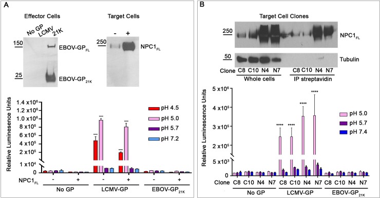
Fig 5. Cells expressing full length NPC1 at the cell surface do not support detectable fusion with cells expressing Ebola GPcl.
(A) Transient NPC1 expression: Effector HEK293T/17 cells were transfected to express the indicated (or No) GP and DSP1-7. Target cells were transfected to express (+) or not (-) full-length NPC1 and DSP8-11. Effector and target cells were analyzed, respectively, for surface exposed EBOV-GP1 and NPC1 following surface biotinylation, avidin precipitation and blotting for EBOV GP or NPC1. Cocultures of effector and target cells were then treated for CCF and analyzed as in Fig 1A. (B) Stable NPC1 expression: (Top) Selected clones (C, control or N, expressing full-length NPC1) were analyzed for NPC1 in whole cell lysates by western blots (left set) and for cell surface exposed NPC1 as in A. Blotting for beta-tubulin was used as a control for protein loading (left set) and cell intactness (right set). Parallel sets of cells depicted in the gels were then used as targets in CCF experiments with effector cells expressing the indicated (or No) GP. Cocultured effector and target cells were then processed as in the graph shown in panel A. Results represent mean +/- SD of triplicate samples from one of two experiments with similar results. Statistical analyses are based on two-way ANOVA tests; each sample was compared to the respective No GP sample ****p < 0.0001.

