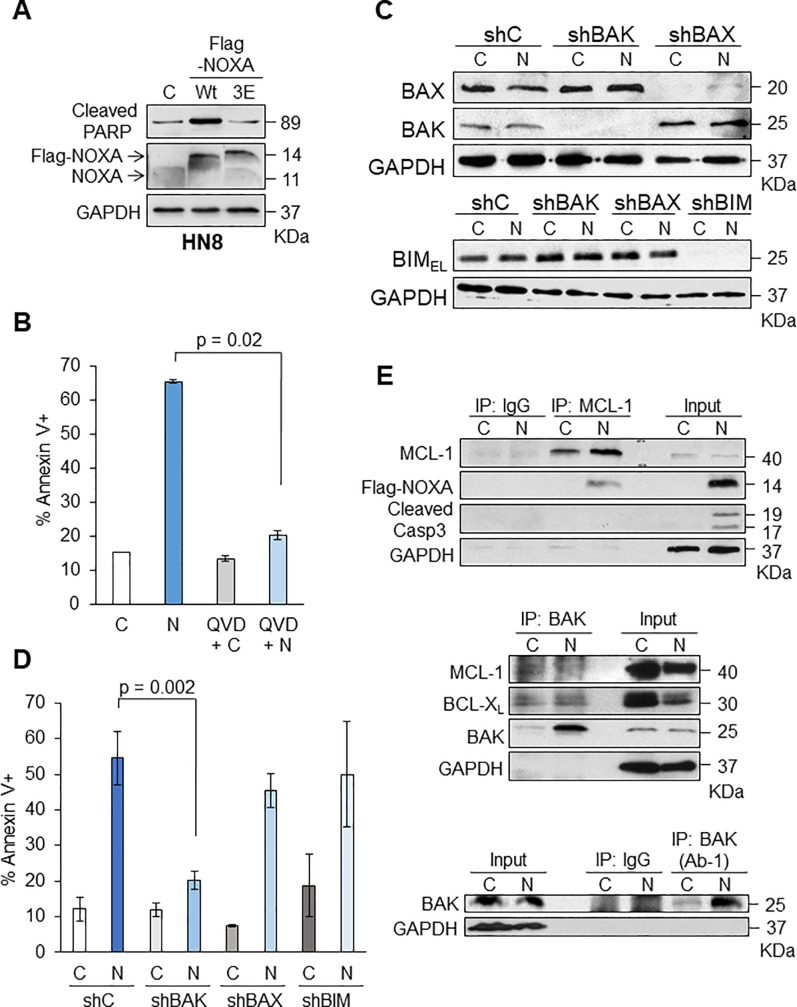
Fig 1. Ectopic NOXA expression alone induces apoptosis through BAK in HN8 cells.
(A) HN8 cells were infected with lentivirus-encoding Flag-tagged NOXA wild-type (Wt), NOXA 3E or vector alone as control (C) for 24 h. Equal amounts of total extracts were subjected to Western blot analysis with the indicated antibodies. (B) NOXA alone induces apoptosis in HN8. Cells were treated with vector-control adenoviruses (Ad-Con, C), NOXA expressing adenoviruses (Ad-NOXA, N), Q-VD-OPH with Ad-Con (QVD+C), and Q-VD-OPH with Ad-NOXA (QVD+N). After 24 h, cells were analyzed with Annexin V-PI staining followed by FACS to determine the amount of apoptosis (N = 3). Values represent the means ± S.D. (C) BAK is mainly contributing to NOXA-induced cell death in HN8 cells. Lentiviruses encoding short-hairpin BAK, (shBAK), BAX (shBAX), BIM (shBIM), and non-targeting control (shC) were infected in HN8 cells and stable cell lines were established with puromycin selection. Cells were then treated with Ad-Con (C) and Ad-NOXA (N) for 16 h followed by Western blot analyses. (D) The cells in (C) were treated with Ad-Con and Ad-NOXA for 24 h followed by FACS analyses to determine total amount of apoptosis (N = 3). Values represent the means ± S.D. (E) NOXA binds to MCL-1 followed by BAK activation in HN8 cells. HN8 cells were treated with Ad-Con (C) and Ad-NOXA (N) for 16 h. Equal amounts of total extracts were incubated with IgG, anti-MCL-1 (top), anti-BAK (middle), or conformation-specific anti-BAK (bottom) antibodies. The input represents 20/500 of the immunoprecipitated lysates. Top: Immunoprecipitation with anti-MCL-1 followed by Western blots with the indicated antibodies. Middle: Immunoprecipitation with anti-BAK followed by Western blots with the indicated antibodies. Bottom: Immunoprecipitation with anti-BAK that detects a conformational change for BAK activation.

