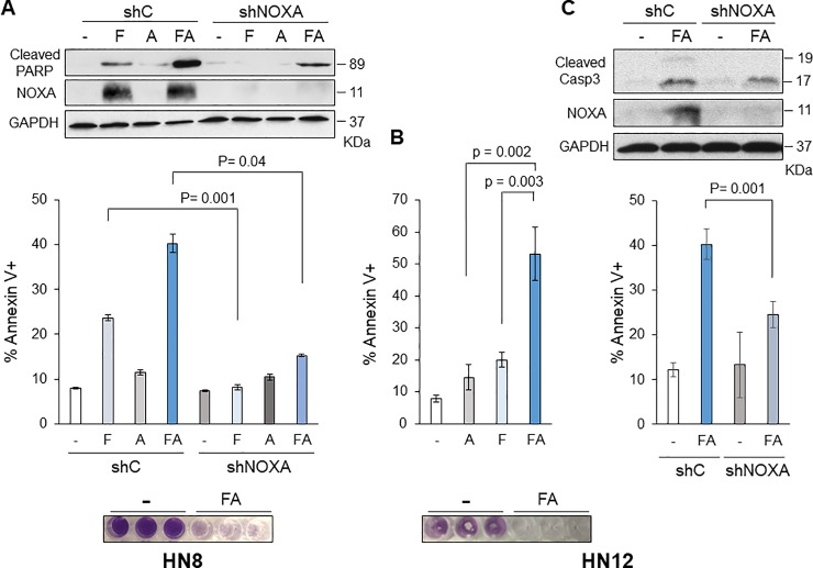
Fig 3. Combination of fenretinide and ABT-263 efficiently induces apoptosis in HN8 and HN12 cells.
(A) Top: HN8 cells were infected with lentivirus-encoding shRNA for non-targeting control (shC) or NOXA (shNOXA). Cells were treated with fenretinide (F; 10 μM) and/or ABT-263 (A; 1 μM) for 16 h. Equal amounts of the total extracts were used for Western blot analysis with the indicated antibodies. Middle: The amount of apoptosis was determined by Annexin V-PI staining followed by FACS analyses (N = 3). Values represent the means ± S.D. Bottom: HN8 cells were treated with the same as above for 72 h and stained with crystal violet. (B) HN12 cells were treated and analyzed as (A). Values represent the means ± S.D. for five independent experiments. (C) Top: HN12 cells were infected with lentivirus-encoding shRNA for non-targeting control (shC) or NOXA (shNOXA). Cells were treated with fenretinide (F; 10 μM) and ABT-263 (A; 1 μM) for 16 h. Equal amounts of the total extracts were used for Western blot analysis with the indicated antibodies. Bottom: The amount of apoptosis was determined by Annexin V-PI staining followed by FACS analyses (N = 4). Values represent the means ± S.D.

