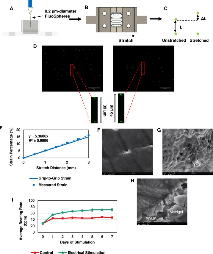
Fig 2. Confirmation of effectiveness of cyclic stretching and electrical stimulation setups.
Fluorescent microspheres were used to measure and standardize the stretching characteristics of the PDMS mold. (A) Microspheres were loaded within each channel and allowed to settle to the bottom, (B) the mold was then attached to the stretching blocks and statically stretched to distances ranging from 0 to 3 mm (based on the actuator distance), and (C) two aligned spheres were chosen to measure the change in distance between them at each increment along with the subsequent strain. (D) Images of the fluorescent spheres at baseline and 3 mm. Scale bar: 100 μm. (E) The generated standard curve from data collected compared to the grip-to-grip strain values. SEM image of (F) only PDMS, (G) PDMS + Matrigel, and (H) combination image of PDMS surface and Matrigel coating. (I) Average beating rate measurement over 7 days of electrical stimulation. These results indicate that each of these stimulation methods were effective in providing the desired interventions of electrical and mechanical stimulation on the spheroids.

