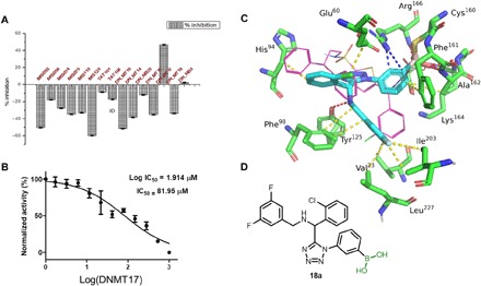
Fig. 6. Covalent inhibition of tuberculosis target MptpB.

(A) Screening of the boronic acid library by a colorimetric enzyme assay. (B) Median inhibitory concentration (IC50) of compound 18a. (C and D) Modeling of compound 18a into MptpB [Protein Data Bank (PDB) ID: 2OZ5], where it forms a covalent adduct with active-site cysteine. Van der Waals interactions, hydrogen bonding, and cation-π interactions are indicated by yellow, red, and blue dotted lines, respectively.
