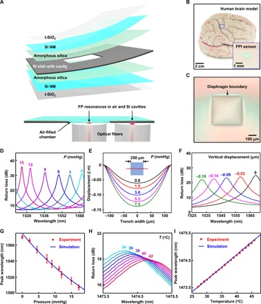
Fig. 1. Materials and designs for bioresorbable FPI-based pressure and temperature sensors.

(A) Schematic illustration of a bioresorbable FPI pressure and temperature sensor composed of a thermally grown silicon dioxide (t-SiO2) encapsulation layer, a Si NM, amorphous silica adhesion layer, and a silicon slab. The layers of t-SiO2 and Si NM serve as pressure-sensitive diaphragms that seal an air chamber formed by bonding with a silicon slab with a feature of relief etched onto its surface. The bottom image shows a cross-sectional view of a sensor integrated with two optical fibers that deliver light to diaphragm and nondiaphragm regions of the device, thereby enabling pressure and temperature sensing, respectively. (B) Photograph of a device placed on an adult brain model. The inset shows a magnified view. (C) Optical micrograph of the top diaphragm. (D) Optical spectra collected from a bioresorbable FPI pressure sensor immersed in PBS (pH 7.4) at room temperature under different pressures. The FP resonance peak associated with the air cavity shifts to the blue with increasing pressure. (E) 3D-FEA of the vertical displacement of the diaphragm along the midsection (red dotted line) at different pressures. (F) CEM simulations of optical spectra obtained at different displacements of the diaphragm. (G) Calibration curve for the FPI pressure sensor (red) compared with simulation results (blue) obtained in (C) and (F). (H) Optical spectra collected from a bioresorbable FPI temperature sensor immersed in PBS at varying temperatures. The FP resonance peak associated with silicon shifts to the red with increasing temperature. (I) Calibration curve for the FPI temperature sensor (red) compared with optical simulation results (blue). Circles and error bars in (G) and (I) indicate means ± SEM for three measurements.
