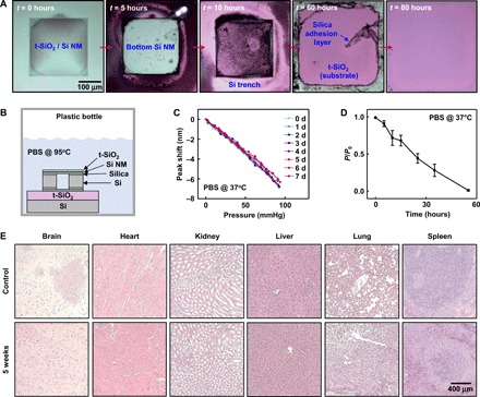
Fig. 4. Kinetics of dissolution and histological studies of bioresorbable optical sensors and fibers.

(A) Optical micrographs at various stages of accelerated dissolution of a bioresorbable FPI sensor due to immersion in PBS (pH 7.4) at 95°C. (B) Schematic illustration of the setup used for the dissolution experiment in (A). (C) Pressure calibration curves obtained from a bioresorbable FPI pressure sensor (t-SiO2 thickness, 1 μm; air chamber thickness, 100 μm), integrated with a commercial optical fiber, soaked in PBS at 37°C for 8 days. The minimal changes in pressure sensitivity provide evidence for the effectiveness of t-SiO2 as a biofluid barrier. (D) Loss of transmission efficiency of the PLGA fiber over time due to immersion in PBS at 37°C. Circles and error bars indicate means ± SEM for four measurements. (E) Representative histological results of the brain, heart, kidney, liver, lung, and spleen organs explanted from a control mouse and a mouse with a bioresorbable FPI sensor implanted in the brain for 5 weeks. The results indicate an absence of abnormalities such as necrosis or inflammation.
