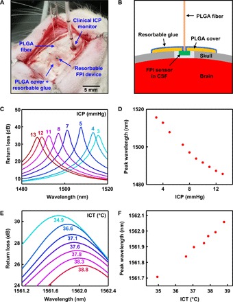
Fig. 5. Acute monitoring of ICT and ICP in rats.

(A) Photograph of a bioresorbable FPI sensor implanted in the intracranial space of a rat for monitoring ICT and ICP. A thin film of PLGA, with a hole in the middle for the fiber, and bioresorbable glue seal the cranial defect to form an airtight environment for accurate pressure sensing. A clinical fiber-optic ICP monitor, implanted nearby, provides reference data. (B) Cross-sectional schematic illustration of the device setup during animal testing. Cutting through the dura and exposing the device to CSF allows assessment of the ICP. (C and D) Optical spectra and pressure calibration curve obtained from a bioresorbable FPI pressure sensor, calibrated against the clinical ICP monitor. Contracting the flank of the rat temporarily raises the ICP by up to ~10 mmHg from the initial level. (E and F) Optical spectra and temperature calibration curve obtained from a bioresorbable FPI temperature sensor, calibrated against a commercial thermistor implanted nearby. The electrical heating blanket wrapped around the body of the rat gradually increased the ICT. (Photo credit for part A: Jiho Shin, University of Illinois at Urbana-Champaign.)
