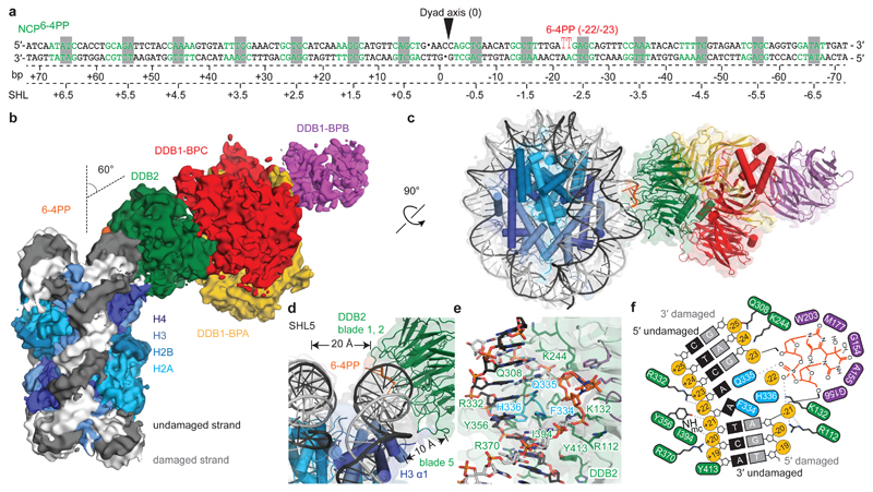
Figure 1. Cryo-EM structure of the NCP6-4PP-UV-DDB complex.
a, DNA sequence with a 6-4PP placed -22/-23 bp from the dyad axis. b, NCP6-4PP-UV-DDB cryo-EM map at 4.3 Å resolution. c, d, NCP6-4PP-UV-DDB domain architecture (cartoon). DDB2 blade 1 (residues 150-156), blade 2 (residues 195-200) and blade 3 (residues 360-370). e, Close-up of the DDB2 β-hairpin loop (cyan) and 6-4PP lesion (orange), with damaged (light grey) and undamaged (dark grey) DNA strands shown. c, d, e Surface depiction of the 4.3 Å resolution cryo-EM map (Extended Data Fig. 4a, b). f, Schematic representation.

