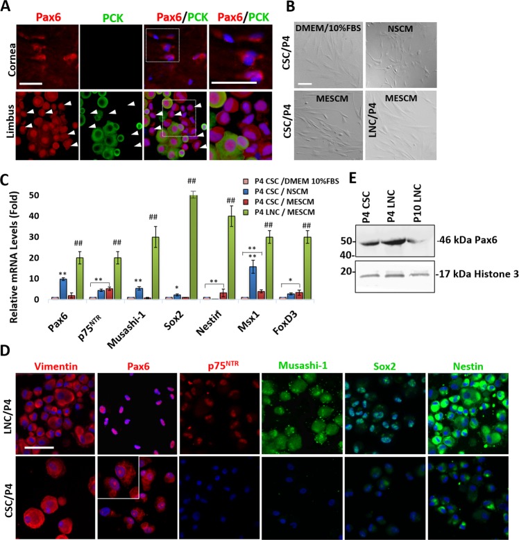
Figure 1.
Unique Nuclear 46 kDa Pax6 in Limbal Niche Cells (LNC). Freshly isolated PCK (−) LNC (arrows) and PCK (+) limbal epithelial cells from the limbal tissue exhibited positive nuclear staining of Pax6 while freshly isolated PCK (−) CSC from epithelially denuded corneal stroma exhibited cytoplasmic staining of Pax6 (A). LNC and CSC were expanded in the same manner on coated Matrigel in MESCM up to passage 4 (P4) while CSC were also cultured on plastic in neural stem cell medium (NSCM) or DMEM/10% FBS. Comparison was made on cell morphology by phase microscopy on day 6 (B). Transcript expression by RT-qPCR of neural crest markers (Pax6, p75NTR, Musashi-1, Sox2, Nestin, Msx1, and FoxD3) in P4 LNC was compared to that of P4 CSC under the identical culture condition (C, ##p < 0.05, n = 3). P4 CSC in MESCM was further compared to P4 CSC expanded in NSCM using the control P4 CSC in DMEM/10%FBS set as 1 (C, *p < 0.1; **p < 0.05, n = 3). Immunofluorescence staining showed the cytolocalization of vimentin (red), Pax6 (red), p75NTR (red), Musashi-1 (green), Sox2 (green) and Nestin (green) in P4 LNC and P4 CSC on coated Matrigel in MESCM (D, nuclear counterstaining by Hoechst 33342). Scale bars = 100 µm in (A,B,D). Protein expression of Pax6 from P4 CSC, P4 LNC and P10 LNC were confirmed by Western blot using Histone 3 as a loading control (E).

