Figure 3.
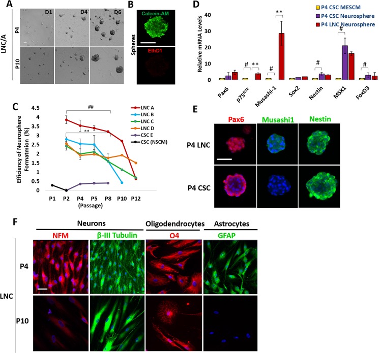
Neural Potential of LNC and CSC Declines after Serial Passages. For each passage, 5 × 10³/cm2 LNC cells were seeded on 12 well plate coated with poly-HEMA in NSCM neurosphere medium to generate neurospheres for 6 days (A, representative images from P4 and P10). Live and dead assay showed the sphere formed by P4 LNC was alive on day 6 (green) without dead cells (red) (B). The neurosphere-forming efficiency (%) was measured from LNC expanded from four different limbal regions and was compared with that of CSC region at each passage (C, ##p = 0.001; **p = 0.001). The transcript level of neural crest markers such as Pax6, p75NTR, Musashi-1, Sox2, Nestin, Msx1, and FoxD3 in neurospheres formed by P4 CSC was compared with those by P4 LNC or P4 CSC seeded on coated Matrigel in MESCM which the transcript expression was set as 1 (D, **p = 0.0001; #p = 0.001, n = 3, respectively). Immunofluorescence staining showed cytolocalization of Pax6 (red), Musashi-1 (green) and Nestin (green) in neurospheres derived from P4 CSC and P4 LNC (E). P4 or P10 LNC were assessed for their potential of differentiation into neurons, oligodendrocytes, and astrocytes by immunofluorescence staining of neurofilament M (NFM, red) and β-III tubulin (green), O4 (red), and Glial fibrillary acidic protein (GFAP, green), respectively (F). Nuclear counterstaining by Hoechst 33342 in (E,F). Scale bars = 50 µm in (A,F), 200 µm in (B), and 100 µm in (E).

