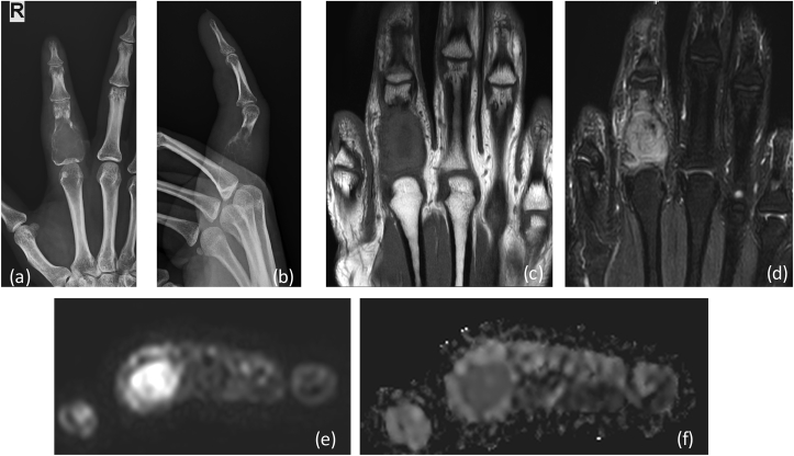
Fig. 8a.
X-ray (a) AP (b) lateral reveals a large expansile lytic lesion on in proximal shaft of proximal phalanx of 4th finger extending up to the articular margin: GCT. No matrix mineralization noted. MRI (c) T1W coronal, (d) T2 FAT SAT Images reveal heterogenous intensity mass lesion in proximal phalanx with thinning of overlying cortex. DWI (e) images show restriction in the soft tissue with reversal of signal on ADC (f).

