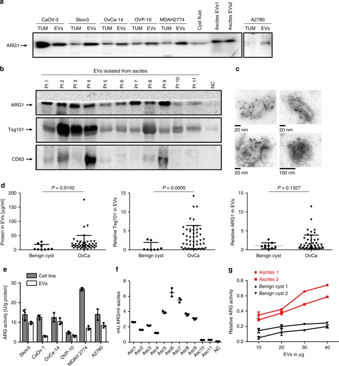
Fig. 2.
OvCa-derived EVs contain enzymatically active ARG1. a ARG1 content in EVs and the parental OvCa cell lines lysates (TUM) as well as in EVs isolated from the cyst fluid and ascites fluid of two OvCa patients (EV1, EV2) determined by Western blotting. Equal amounts of protein (30 μg) were loaded per lane. b Representative Western blot for ARG1 and typical exosomal markers in EVs isolated from ascites of n = 11 OvCa patients. EVs isolated from ovarian fluid served as normal control (NC). Equal amounts of protein (30 μg) were loaded per lane. c Representative TEM images of whole-mounted OvCa patient-derived tEVs. Dots indicate immunogold (6 nm gold particles) labeling of ARG1. d Protein concentration measured with BCA assay, Tsg101 and ARG1 levels in EVs isolated from 2 ml of ascites of OvCa patients (n = 47–50) and from 2 ml of benign cysts fluid (n = 7–9). Relative Tsg101 and ARG1 content was determined by densitometric analysis of Western blots. Data refers to means ± SD, P values were calculated with unpaired t-test with Welch’s correction. e Arginase activity in OvCa cell line lysates and in the corresponding EVs determined by measuring ʟ-arginine conversion to urea in a colorimetric assay. Data show means ± SD, n = 3. f Arginase activity in EVs isolated from n = 11 OvCa patients’ ascites (Asc) and benign cyst fluid (NC) calculated per ml of starting fluid volume. Data show means ± SD, n = 3. g Arginase activity as a function of amount of EVs in n = 2 OvCa patients ascites-isolated tEVs and EVs isolated from n = 2 benign cyst fluid. Data show means ± SD. Source data for data in panels a, b, d–g are provided as a Source Data file

