Fig. 4.
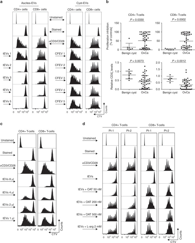
EV-ARG1 is involved in the suppression of T-cell proliferation in vitro. a Representative proliferation histograms of αCD3/αCD28-stimulated CD8+ and CD4+ T cells incubated for 6 days with tumor ascites-derived EVs isolated from n = 5 OvCa patients (left panel, tEVs 1–5) and control EVs (CFEV1–5, right panel), isolated from n = 5 patients with benign cyst of the ovary. b Inhibition of proliferation (upper) and decrease in CD3ζ levels (lower) of peripheral blood CD4+ (left) or CD8+ T-cells (right) by OvCa ascitic fluid-isolated EVs (n = 43–44). Data show means ± SD, P values for OvCa ascites vs. benign cyst fluid-isolated EVs treated group (n = 6–7) were calculated with Mann–Whitney U-test. The amount of added EVs corresponded to 2 ml of starting material a, b. c Representative proliferation histograms of αCD3/αCD28-stimulated CD4+ and CD8+ T-cells incubated with increasing amounts of tEVs, isolated from 2 ml (8 µl), 1 ml (4 µl), 0.5 ml (2 µl), or 0.25 ml (1 µl) of ascites, respectively. d Representative proliferation histograms of peripheral blood CD4+ and CD8+ T-cells incubated for 6 days with tEVs (corresponding to 2 ml of ascites) and indicated concentrations of an arginase inhibitor, OAT-1746. Cells incubated for 6 days with tEVs and 2 mM ʟ-arginine served as a positive control for ʟ-arginine-dependent reversal of T-cells proliferation inhibition. Source data for panel b are provided as a Source Data file

