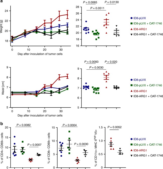
Fig. 7.
ARG1 promotes tumor growth in vivo. C57BL/6 mice were inoculated i.p. with 4 × 10⁶ ID8-VegfA/Defb29 cells transduced with V5-tagged murine ARG1 (ID8-ARG1-V5) or the control vector (ID8-pLVX). a Mice were treated from day 14th after tumor inoculation with OAT-1746 or PBS i.p. twice daily and monitored for tumor development until first mice met the humane endpoint criteria described in the “Methods” section. Increase in mice weight (upper left) and waist circumference (lower left) in time compared to day 0 (day of tumor cells i.p. inoculation) as a measure of ovarian cancer progression/ascites development. Measurements of gained weight (upper right) and percentage of gained waist circumference (lower right) on day 34 after inoculation of tumor cells. Each experimental group consisted of n = 5–9 mice. Data show means ± SE. b Mice were treated from day 14th after tumor inoculation with OAT-1746 ARG inhibitor or PBS i.p. twice daily for consecutive 2 weeks. After 2 weeks of treatment cells from ascites were isolated and analyzed by flow cytometry. Percentage of ascitic activated (identified as CD69+) CD4+ (left panel) or CD8+ T-cells (middle panel), as well as percentage of activated (MHCIIhigh) CD11c+ DCs positive for V5-tag (right panel) on day 28th after inoculation of tumor cells. Each experimental group consisted of n = 3–6 mice. Data show means ± SD; P values were calculated with unpaired t-test. Source data are provided as a Source Data file

