Fig. 2.
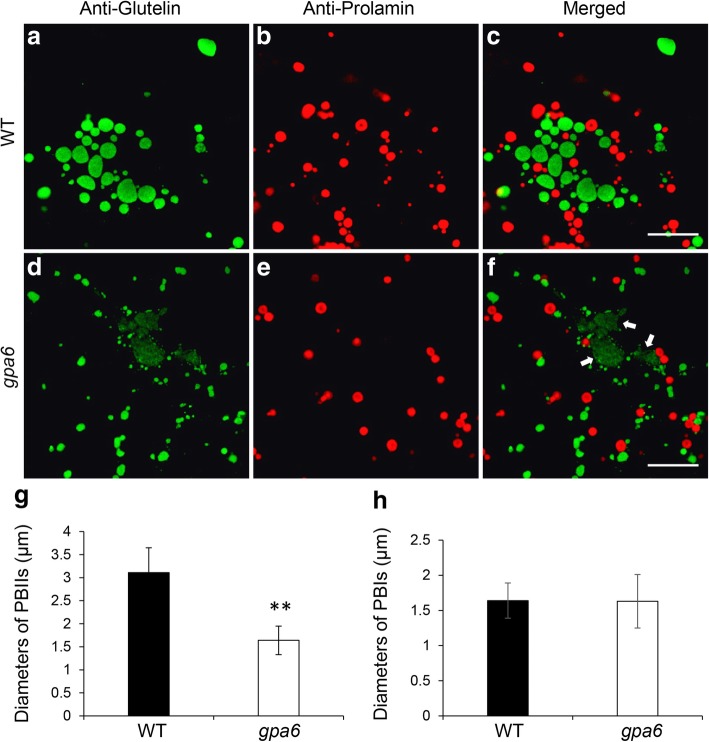
Immunofluorescence microscopy of protein bodies in the subaleurone cells of the wild type and gpa6 mutant. a to f Immunofluorescence microscopy images of storage proteins in wild-type (a-c) and gpa6 (d-f) 12 DAF seeds. a, d Secondary antibodies conjugated with Alexa fluor 488 (green) were used to trace the antigens recognized by the anti-glutelin antibodies. (b, e) Secondary antibodies conjugated with Alexa fluor 555 (red) were used to trace the antigens recognized by the anti-prolamin antibodies. (c, f) Merged images. White arrows in (f) indicate the PMB structures. Bars = 10 μm (a-f). (g) and (h) Statistical analysis of the diameters of PBIIs (g) and PBIs (h). Values are means ± SD. **P < 0.01 (n > 350, Student’s t test)

