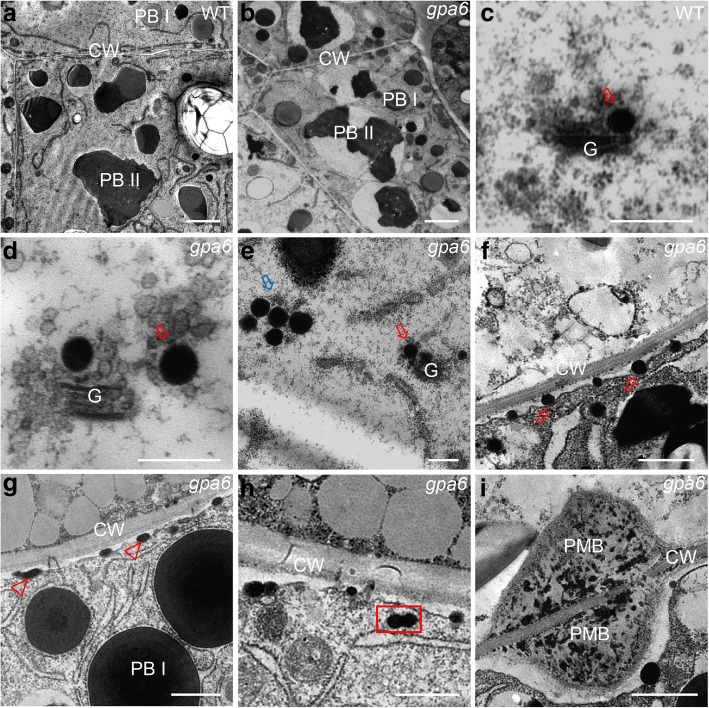
Fig. 3.
Ultrastructure of subaleurone cells of developing endosperm of the wild type and gpa6 mutant. a and b Two types of protein bodies were observed in wild-type (a) and gpa6 mutant (b) endosperm. Bars = 2 μm. CW, cell wall. c Wild type. Bars = 400 nm. d DVs bud off from the Golgi in the gpa6 mutant. Red Arrow indicates enlarged DVs. Bars = 400 nm. e Large clusters of DVs (blue arrow) in the gpa6 mutant. Bars = 400 nm. f and g Electron micrographs showing that DVs can fuse with the PM (f) and expel their contents into the apoplast forming oval-shaped structures (Arrowheads) (g) in the gpa6 mutant. Bars = 1 μm. h Two DVs are fused with each other (rectangular box) in the gpa6 mutant. Bars = 1 μm. (i) The PMB structures in the gpa6 mutant. Bars = 1 μm

