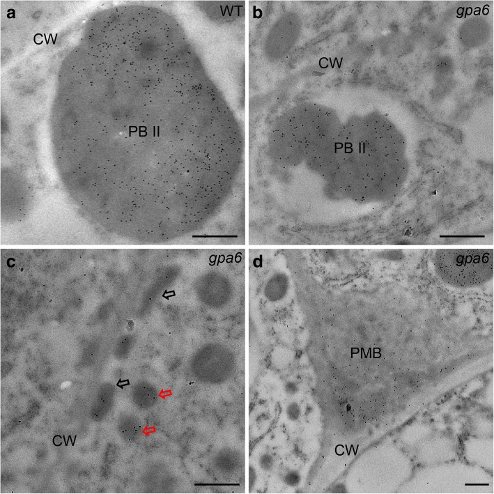
Fig. 4.
Immunoelectron microscopy localization of glutelins in rice endosperm cells. a Glutelins were accumulated in PBIIs in the wild-type endosperm cells. CW, cell walls. Bars = 500 nm. b Size-reduced PBII containing glutelins. Bars = 500 nm. c Glutelins in DVs (red arrows) and oval-shaped structures (black arrows). Bars = 500 nm. d Glutelins in the PMBs. Bars = 500 nm. 10-nm gold particle conjugated secondary antibodies were used in (a–d)

