Fig. 7.
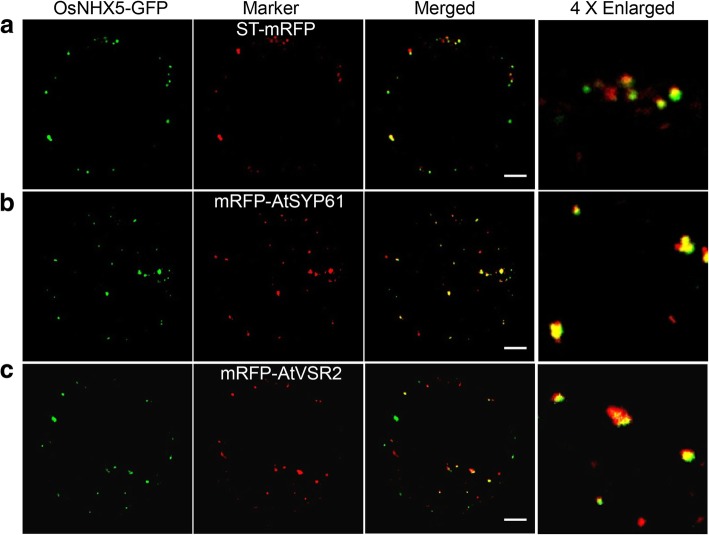
Subcellular Localization of OsNHX5 in N. benthamiana protoplasts. a to c Confocal microscopy images showing that OsNHX5-GFP is localized as punctate signals in the cytosol and its distribution partially overlaps with the markers for Golgi (ST-mRFP [a]), TGN (mRFP-SYP61 [b]) and PVC (mRFP-VSR2 [c]). Bars = 10 μm (a-c)

