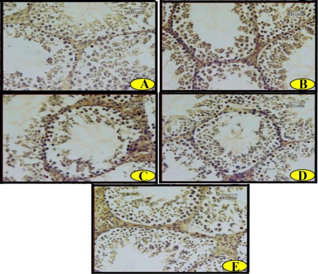
Figure 3.
Photomicrographs of apoptotic cells in the testis of (A) control, (B) healthy animals that received troxerutin, (C) diabetic animals, (D) diabetic animals that received troxerutin and (E) diabetic animals that received insulin. Brown-yellow dots display the positive (apoptotic) cells. (Terminal deoxynucleotidyl transferase dUTP nick end labeling (TUNEL); X400)

