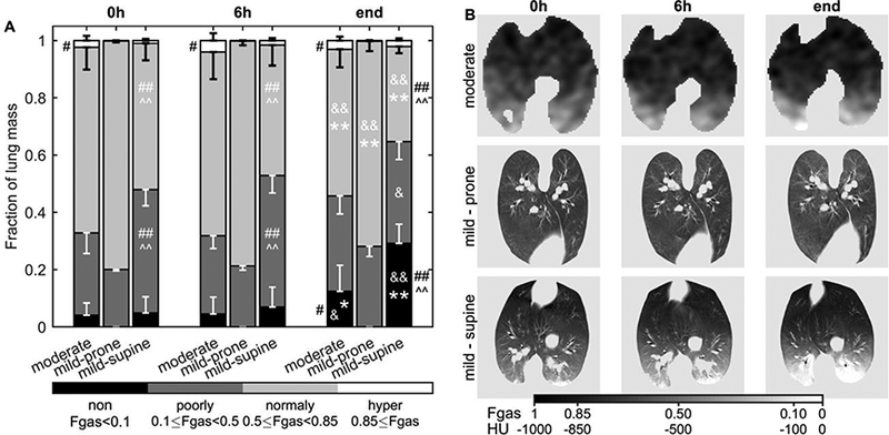
Figure 1-.

Lung aeration in sheep mechanically ventilated with low tidal volume and low to moderate positive and-expiratory pressure for 20–24h. After the first measurement, intravenous infusion of LPS was started to generate moderate (10 ng/kg/min) or mild (2.5 ng/kg/min) systemic endotoxemia. Aeration was quantified as the gas fraction (Fgas) in each voxel of transmission (moderate group) and computed tomography (mild groups) scans during tidal ventilation. (A) Non- (black), poorly- (dark grey), normally- (light grey) and hyper-aerated (white) compartments are expressed as a fraction of total lung mass. Irrespective of the endotoxin dose, non-aerated regions increased after 20h in supine, but not in prone animals. For supine sheep, the spatial distribution of aeration followed a gravitational gradient decreasing toward dorsal regions (B). Aeration level at 24h * vs 0h and & vs 6h for the same group; Λ vs moderate and # vs mild-prone for the same aeration compartment; one symbol p<0.05, two symbols p<0.001.
