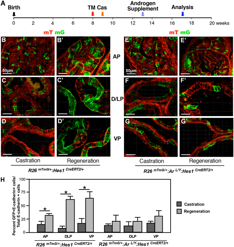
Figure 6. Deletion of AR expression in Hes1-expressing cells impairs prostate regeneration.
(A) Schematic of experimental timeline including tamoxifen induction, castration, regeneration and analysis of prostate tissues. Cas: castration. (B-D) mTmG analysis of different prostatic lobes in R26mTmG/+: Hes1CreERT2/+ mouse tissues prior to or post castration as labeled above. AP: anterior prostate, DLP: dorsolateral prostate, VP: ventral prostate. (E-G) Similar analyses were conducted in different prostatic lobes of R26mTmG/+: ArL/Y: Hes1CreERT2/+ mice. (H) Quantification of GFP and E-cadherin co-expressing cells in prostate lobes isolated from the indicated mice. Please also see Supplemental Table 8 in the Supplemental data file. Scale bars: 50 μm. Results are mean +/− SD of at least three independent samples per group. *P<0.05.

