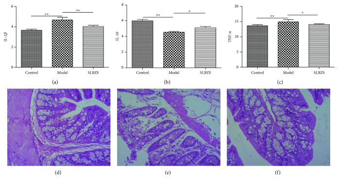
Figure 6.
IL-1β (a), IL-10 (b), and TNF-α (c) can be significantly improved by SLBZS. ∗ P < 0.05; ∗∗ P < 0.01. Histological changes of the colon in the control group (d), the model group (e), and the SLBZS group (f). The colon in the control group (d) presented the normal histological feature. In the model group (e), there was intestinal inflammatory cell infiltration, intestinal villus epithelial cell degeneration, necrosis, and shedding. In the SLBZS group (f), the pathological change was significantly reduced. HE staining (×100).

