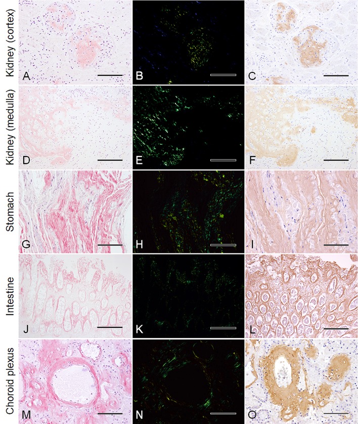
Fig. 1.
Histologic features of systemic amyloidosis in Stejneger’s beaked whales (Mesoplodon stejnegeri). Columns: left, Congo red; middle, Congo red under crossed polars; right, immunohistochemistry for amyloid A with Mayer’s hematoxylin counterstain. (A)–(C) Cortex of kidney (serial sections). Segmental amyloid deposits expand the glomeruli (SNH18001). Bar=100 µm. (D)–(F) Medulla of kidney (serial sections). Marked amyloid deposits in the tubular basement membrane and interstitium at the renal papilla (SNH17015). Bar=200 µm. (G)−(I) Main stomach. Prominent amyloid deposits in the interstitium of the lamina propria (SNH18001). Bar=100 µm. (J)–(L) Intestine. Striking amyloid deposits with a periglandular pattern and at the tip of the lamina propria (SNH17015). Bar=200 µm. (M)–(O) Choroid plexus. Marked amyloid deposits expanding the vascular walls while nodular deposits intersperse within the interstitium (SNH17015). Bar=100 µm.

