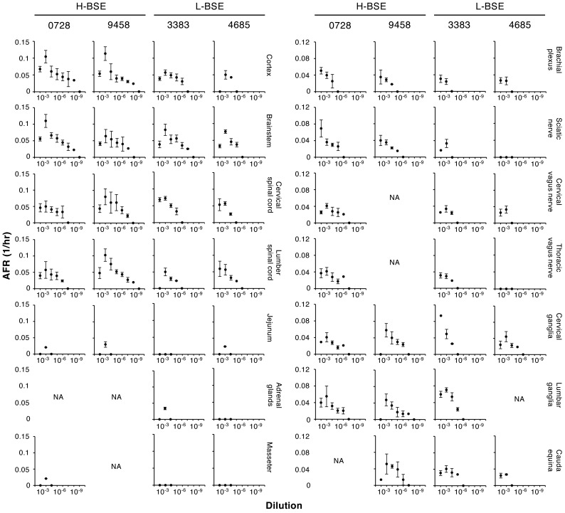
Fig. 2.
Detection of seeding activities in tissues of cattle intracerebrally inoculated with H- and L-BSE. Tissues from two cattle intracerebrally inoculated with H-BSE (IDs: 0728 and 9458) or L-BSE (IDs: 3383 and 4685) were subjected to RT-QuIC. Graphs show AFRs (mean ± standard deviations from a total of eight wells from the two independent experiments [n=4 in each experiment]). Tissues positive for H-BSE and/or L-BSE are indicated. Y-axes show AFRs (1/hr) whereas x-axes show the dilution of tissue homogenates. NA: tissues were not available.

