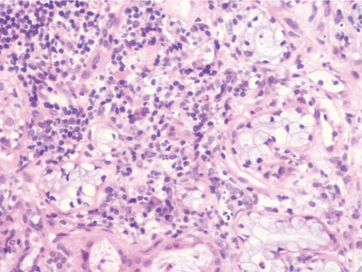
Fig. 3.
Histopathological section of the submandibular salivary glands. Tissue samples obtained from the submandibular salivary glands by Tru-cut biopsy on Day 1 were subjected to hematoxylin-eosin staining according to a conventional method. The section showed mild to moderate lymphoplasmacytic infiltration with a small number of neutrophils in the stroma. Pyknotic or degenerated epithelial cells were occasionally seen. There was no evidence of neoplastic proliferation or infectious agents.

