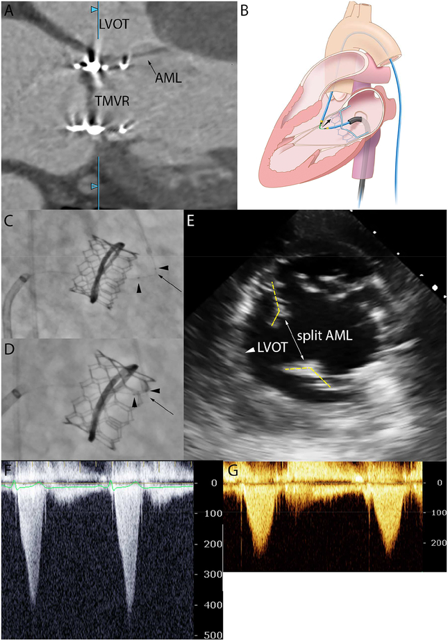
Figure: The “rescue” LAMPOON procedure.
[A] Cardiac-gated CT shows SAM after TMVR. [B] Illustration of concept: a kinked guidewire lacerates the AML in the direction of the black arrow. [C] Fluoroscopy in RAO with kinked mid-shaft of the Astato guidewire (black arrow) sheathed in two microcatheters (black arrow heads) guided by a transseptal sheath and transfemoral catheter. [D] The guidewire is electrified and pulled to lacerate the native AML. [E] Mid-gastric transesophageal view confirming a split AML parted away from the LVOT. [F-G] Severe LVOT gradient is treated.

