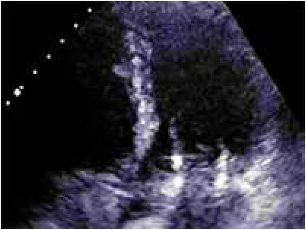
VIDEO LEGEND.
Transthoracic echocardiogram and cardiac-gated CT images show SAM after TMVR. Fluoroscopy and transesophageal echocardiography shows the kinked guidewire between the transseptal sheath and transfemoral guiding catheter being retracted during radiofrequency energy application. The guidewire can be seen advancing towards the Sapien3 valve frame and microbubbles are seen from guidewire electrification. Transesophageal echocardiogram confirms midline laceration of the anterior mitral leaflet in front of the LVOT.

