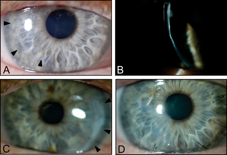
Figure 2:
Slit-lamp photos of the proband’s right cornea demonstrating the location of prior temporal paracentral perforations (arrowheads) (A) and diffuse thinning (B). Images obtained 4 months after epi-off CXL. Slit-lamp photos of the proband’s left cornea demonstrating the location of prior temporal paracentral spontaneous perforation that developed 8 days after epi-on CXL (arrowheads) (C). Three months after the third spontaneous perforation in the right cornea, which occurred 3 years after CXL, sutures are present in the location of the corneal perforation (D).

