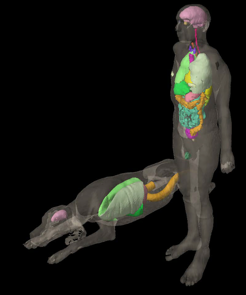
Figure 4:
The UF canine anatomic phantom (prone) and ICRP 110 reference male phantom, with select organ contours visualized within the external beam planning module of the Varian Eclipse TPS. The voxel resolutions (x,y,z) were (0.20 cm, 0.20 cm, 0.20 cm ) and (0.2137 cm, 0.2137 cm, 0.80 cm) for the UF canine and ICRP 110 phantom, respectively.

