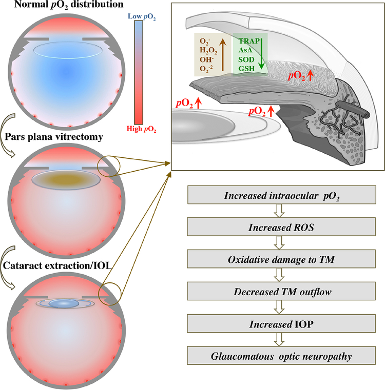
Figure 5.
Proposed mechanism for development of post-vitrectomy glaucoma. Upper left image: Low levels of pO2 in vitreous cavity, surrounding clear lens and in anterior chamber (AC) angle. Oxygen diffuses across cornea into the AC and from retinal blood vessels into the vitreous cavity. Center left image: Following pars plana vitrectomy surgery, pO2 increases in the vitreous cavity, around lens, in AC angle and posterior chamber with development of nuclear sclerotic cataract. Lower left image: Following cataract extraction and intraocular lens implantation (IOL), pO2 increases in the AC angle, posterior chamber and mid-AC. Upper right image: Expanded view of AC angle illustrating alterations in aqueous humor including increased pO2 surrounding lens implant and in AC angle leading to increased reactive oxygen species formation (brown; ROS) and decreased antioxidants (green). Lower right image: Proposed cascade of events following vitrectomy and IOL implantation ultimately leading to increased intraocular pressure (IOP) and risk of glaucoma.
O2− = superoxide anion, H2O2 = hydrogen peroxide, OH− = hydroxyl ion, O2−2 = peroxide, TRAP = total reactive antioxidant potential, AsA = ascorbate, SOD= superoxide dismutase, GSH= glutathione, TM = trabecular meshwork

