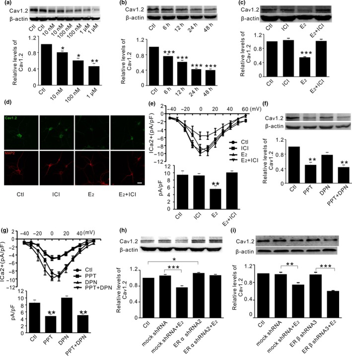
Figure 1.

Estrogen decreases Cav1.2 protein through ERα in primary cortical neurons. (a) Representative Western blots (top) and quantification (bottom) of Cav1.2 in primary cortical neurons in the absence (Ctl) and presence of estrogen at 10, 100 nM, and 1 μM, respectively; n = 4. (b) Representative Western blots (top) and quantification (bottom) of Cav1.2 in cortical neurons incubated with estrogen (100 nM) at different times; n = 3. (c) Representative Western blots (top) and quantification (bottom) of Cav1.2 in cortical neurons treated with DMSO (Ctl), the ER antagonist ICI182780 (1 μM, ICI), estrogen (100 nM, E2), and E2 with ICI, respectively n = 4. (d) Representative immunofluorescent images of Cav1.2 (green) relative to neuronal marker MAP2 (red), in cortical neurons treated with DMSO (Ctl), ICI, E2, and E2 with ICI for 24 hr (n = 3). Scale bar, 10 μm. (e) I‐V plots (top) and quantifications (bottom) of calcium mediated current (ICa2+) density (pA/pF) in primary cortical neurons treated with DMSO (Ctl, n = 15), ICI (n = 9), E2 (n = 9), and E2 with ICI (n = 11) for 24 hr. (f) Representative Western blots (top) and quantification (bottom) of Cav1.2 in primary cortical neurons treated with ERα agonist PPT (10 nM), ERβ agonist DPN (10 nM), and PPT with DPN for 24 hr; n = 6. (g) I‐V plots (top) and quantifications (bottom) of ICa2+ density (pA/pF) in cortical neurons treated with DMSO (Ctl, n = 15), PPT (n = 9), DPN (n = 9), and PPT with DPN (n = 11). (h) Representative Western blots (top) and quantification (bottom) of Cav1.2 in cortical neurons in the absence (Ctl) and presence of mock shRNA, mock shRNA with estrogen (E2, 100 nM), ERα shRNA2, and ERα shRNA2 with E2, respectively (n = 4). (i) Representative Western blots (top) and quantification (bottom) of Cav1.2 in cortical neurons in the absence (Ctl) and presence of mock shRNA, mock shRNA with estrogen (E2, 100 nM), ERβ shRNA3, and ERβ shRNA3 with E2, respectively (n = 3). *p<0.05,**p<0.01,***p<0.001 (ANOVA)
