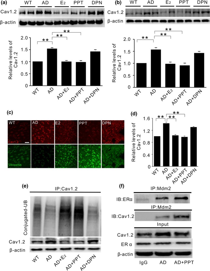
Figure 5.

PPT treatment reduces Cav1.2 protein in ovariectomized APP/PS1 mice. (a and b) Representative Western blots (top) and quantification (bottom) of Cav1.2 in the hippocampus (a) and the cortex (b) of ovariectomized wild‐type mice without any treatment (WT, n = 8), or ovariectomized APP/PS mice treated with vehicle (AD, n = 7), 17β‐estradiol (E2, 30 μg/kg, n = 8), PPT (1 mg/kg, n = 8), and DPN (1 mg/kg, n = 7) for two weeks. E2 or PPT replacement attenuates Cav1.2 elevation in OVX APP/PS1 mice. (c) Representative immunofluorescent images from cortical slices probed with anti‐Cav1.2 (red) and anti‐ubiquitin (green) antibodies, in WT or OVX APP/PS1 mice. (d) Quantification of Cav1.2 immunofluorescent density using data related to C. (e) Western blots of ubiquitin (UB) in cortical extracts immunoprecipitated by Cav1.2 antibody, showing that Cav1.2 ubiquitination is significantly increased in APP/PS1 mice treated with E2 or PPT, but not with DPN. (f) Western blots of ERα (top) and Mdm2 (middle) in the hippocampal extracts of OVX APP/PS1 mice immunoprecipitated by Mdm2 antibody. Proteins in input are shown on the bottom. PPT treatment increases Mdm2 association with ERα and Cav1.2. *p<0.05, **p<0.01, ***p<0.001, ANOVA
