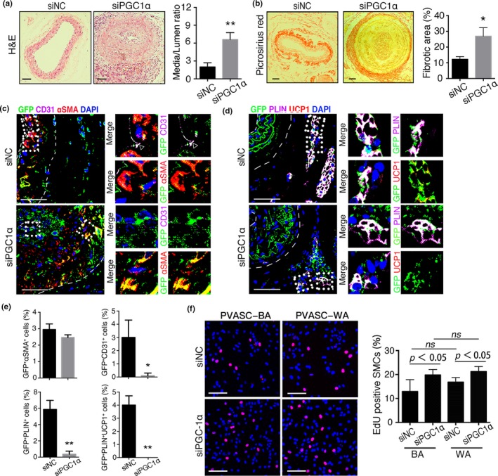
Figure 5.

PGC1α‐knockdown in young PVASCs accelerates neointimal hyperplasia after perivascular delivery. (a,b) H&E and picrosirius red‐stained sections of carotid arteries 28 days after PGC1α‐short heparin RNA (siPGC1α) or negative control (siNC) infected PVASCs delivery to perivascular tissue of injured arteries. (c‐e) The carotid arterial sections were co‐stained for GFP, CD31, SMA (c) and GFP, PLIN, UCP1 (d). Scale bar 50 μm. n = 5 per group. *p < 0.05, **p < 0.01, versus negative control. (f) The proliferation of SMCs cocultured with siPGC1α or siNC‐PVASC‐WA/BA was measured by Edu staining. Red indicated positive staining for Edu. n = 4 independent experiments
