Figure 2.

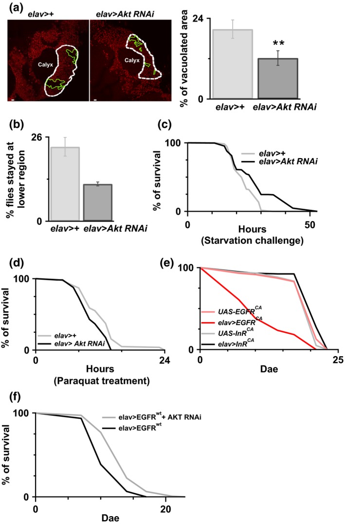
Reduced Akt benefits animal survival. (a) Reduced Akt in neurons by RNAi promotes cell survival in the Kenyon cell body region at the age of 20 days. White dot circle is the place where we used to do the quantification and is above the calyx. Green circles represent the area where there was no clear PI staining, which suggests there were cells lost. Percentage of vacuolated area (i.e. percentage of area circled in green) within calyx was quantified and shown in the right panel. N = 5–7. **p < 0.01. White bar = 2 μm. (b) A better climbing ability was observed in the Akt knocked‐down flies compared to the control flies, without RNAi expression. Less Akt RNAi flies stay at the bottom. (c) Reduced Akt in neurons showed better starvation resistance. The survival rate of elav>Akt RNAi was better than elav>+. p < 0.001 log‐rank analysis. N = 180 for elav>+ and N = 270 for elav>Akt RNAi. (d) Reduced Akt has no any improvement on the oxidative stress. The survival rate between flies, with/without Akt knocked‐down, was similar. N = 180 for elav>+ and N = 110 for elav>Akt RNAi. (e) Overexpression of EGFRCA but not InRCA in the flies promoted animal early death compared to the control flies, flies without any genetic manipulation. N = 120 for each genotype. There was significant lifespan decreasing in the flies with EGFRCA overexpression. p < 0.001 log‐rank analysis. (f) Reduced AKT delays EGFR wt‐induced early death. N = 140–200. p < 0.001 log‐rank analysis
