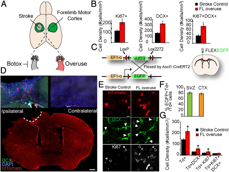
Fig. 1.
Forelimb overuse increases neurogenesis in the peri-infarct cortex after stroke. (A) Schematic demonstration of stroke induction and forelimb overuse. Focal ischemic stroke in the forelimb motor cortex was induced by photothrombosis. To induce overuse of the paretic forelimb, Botox was injected to paralyze the nonparetic forelimb 24 h after stroke. (B) Density of Ki67+, DCX+, and Ki67+DCX+ cells. *P < 0.05. (C) Schematic demonstration of the EF1-α–FLEX–EGFP plasmid and lateral ventricle injection of the lentivirus expressing EF1-α–FLEX–EGFP. Lentivirus EF1-α–FLEX–EGFP was injected into the lateral ventricle of Ascl1–CreERT2::tdTomato animals 7 d before stroke. Tamoxifen was administered daily to the mice from days 2 to 7 after stroke. (D) Low-magnification confocal micrograph of the whole-brain coronal section with tdTomato, DCX, and DAPI 14 d after stroke. (Scale bar: 500 µm.) (E) Confocal micrographs of tdTomato+, DCX+, and Ki67+ cells in the peri-infarct cortex of the Ascl1–CreERT2::tdTomato mice 14 d after stroke. Filled arrowheads, tdTomato+DCX+ cells; open arrowheads, tdTomato+Ki67+ cells. (Scale bar: 5 µm.) (F) Percentage of EGFP+ tdTomato+ cells among total tdTomato+ cells in the SVZ and in the peri-infarct cortex 14 d after stroke. (G) Density of tdTomato+, tdTomato+DCX+, tdTomato+Ki67+, and tdTomato+Ki67+DCX+ cells in the peri-infarct region 14 d after stroke. *P < 0.05.

