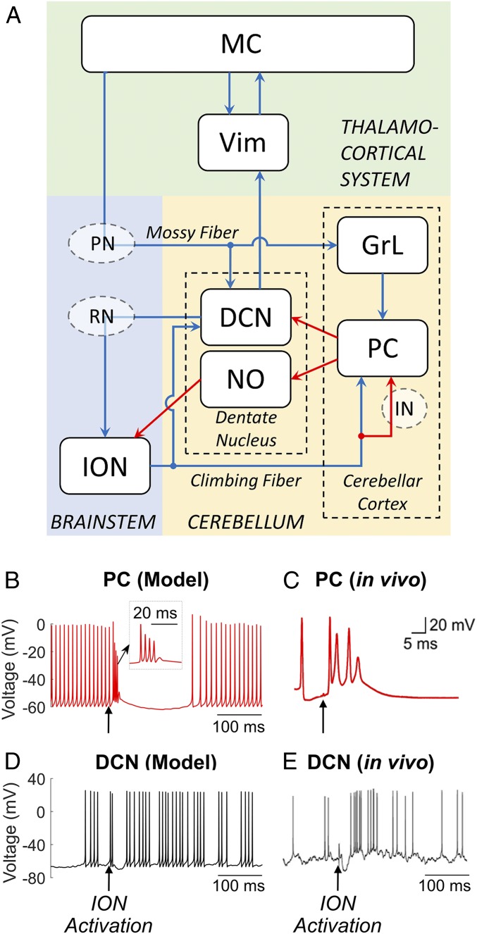
Fig. 1.
(A) Schematic of the CCTC model. Blue arrows, glutamatergic excitatory connections; red arrows, GABAergic inhibitory connections; PC, Purkinje cells; DCN, deep cerebellar neurons; NO, nucleoolivary neurons; ION, inferior olive nucleus; GrL, granular layer; RN, red nucleus; PN, pontine nucleus; IN, cerebellar interneurons; Vim, ventral intermediate nucleus of the thalamus; MC, motor cortex. (B–E) Response of PC and DCN neurons to a depolarizing stimulus applied in the ION in the proposed network model (B and D) and in rodents in vivo (C and E) in normal, tremor-free conditions. A single suprathreshold (10 pA) current pulse (pulse duration of 20 ms) was applied to all ION neurons in our model (black arrows in B and D) and in the inferior olivary nucleus of the rodent (black arrows in C and E) and resulted in a burst of action potentials with amplitude adaptation (i.e., complex spike) in the PC (B and C) and an after-hyperpolarization rebound burst of action potentials in the DCN (D and E). (B, Inset) Zoom-in of the complex spike. Image in C is reprinted from ref. 43, which is licensed under CC BY 3.0. Image in E is reprinted from ref. 45, which is licensed under CC BY 4.0.

