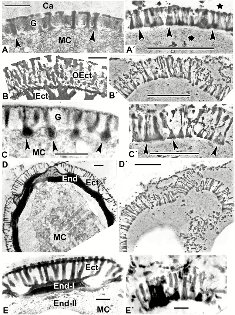
Fig. 2.
Natural exine patterns of a number of species (A–E) and experimental patterns simulated by self-assembly (A′–E′). (A) Columellate ectexine in the process of development at the late tetrad stage in Acer tataricum. Columellae (arrowheads). (Fig. 4a in Gabarayeva et al., 2010a). (A′) Simulation with columellate-like pattern (arrowheads) at the interface between lipidic (asterisk) and aqueous (star) domains. (B) Mature ectexine in Echinops exaltatus with outer ectexine (OEct) consisting of thin columellae (united with each other by fine connections) and of inner ectexine (IEct) of thick columellae. (Fig. 11e in Gabarayeva et al., 2018a). (B′) Simulation mimicking the outer ectexine in Echinops. (C) Late tetrad stage in Cabomba aquatica. Developing columellae have widened ‘feet’ (arrowheads). (Plate III, 9 in Gabarayeva et al., 2003). (C′) Columellae-like simulation; ‘columellae’ have widened ‘feet’ (arrowheads). (D) A free microspore of Borago officinalis with mature columellate ectexine (Ect) and endexine (End). Aperture site is indicated by an arrow. (Fig. 20 in Rowley et al., 1999). (D′) Simulation partly imitating the ectexine in Borago. (E) Magnified interapertural portion of the exine in Borago officinalis with columellate ectexine (Ect) and two-layed endexine (End-I and End-II). (Fig. 21 in Rowley et al., 1999). (E′) Simulation similar to partly distorted interapertural ectexine in Borago. Scale bars: (A, C, E, A′–D′) = 0.5 µm; (B) = 2 µm; (D) = 1 µm; (E′) = 0.1 µm. Ca, callose; G, glycocalyx; MC, microspore cytoplasm.

