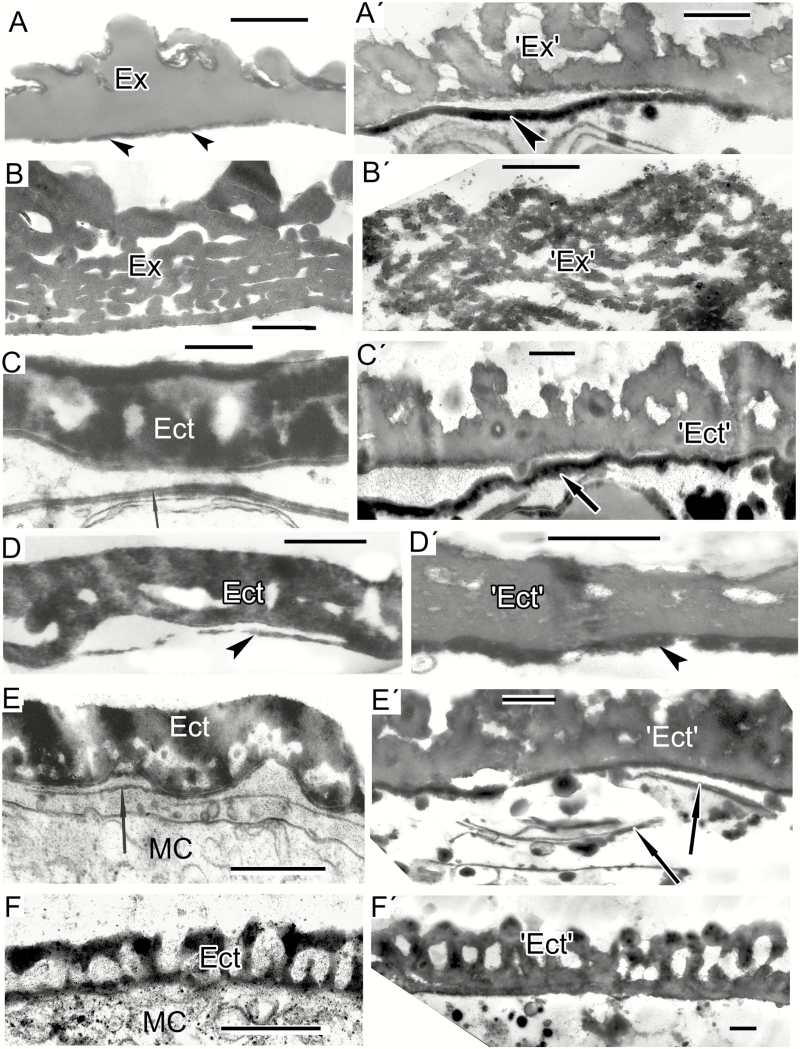
Fig. 5.
Spore and exine patterns of a number of species (A–F) and experimental patterns simulating them by self-assembly (A′–F′). (A, B) Megaspore walls of extinct representatives of the genera Verrucosisporites and Osmundacidites: Verrucosisporites narmianus (A) and Osmundacidites wellmanii (B) (courtesy of V. Tarasevich) and simulations mimicking them (A′, B′). The dark contrast layer in A′ (arrowhead) mimics a probable endospore layer in A (arrowheads). (C) Microspore wall in Magnolia delavayi with ectexine (Ect) and developing endexine (arrow). (Fig. 10A in Gabarayeva and Grigorjeva, 2012). (C′) Model simulating the ectexine and endexine (arrow) of Magnolia microspore wall. (D) Acetolysed pollen wall in Magnolia delavayi, with the endexine indicated by an arrowhead. (Fig. 14E in Gabarayeva and Grigorjeva, 2012). (D′) Simulation resembling the acetolysed pollen wall in Magnolia delavayi. (E) Microspore wall of Michelia fuscata with the ectexine (Ect) and the developing lamellae of the endexine (arrow). (Fig. 10B in Gabarayeva and Grigorjeva, 2012). (E′) Simulation of Michelia microspore wall mimicking ectexine (‘Ect’) and the developing lamellae of the endexine (arrows). (F) Ectexine in Chamaedorea microspadix. (Fig. 7a in Gabarayeva and Grigorjeva, 2010). (F′) Simulation mimicking the ectexine of Chamaedorea. Scale bars: (A, B) = 1 µm; (C–F, A′–F′) = 0.5 µm. Ex, exosporium; MC, microspore cytoplasm.

