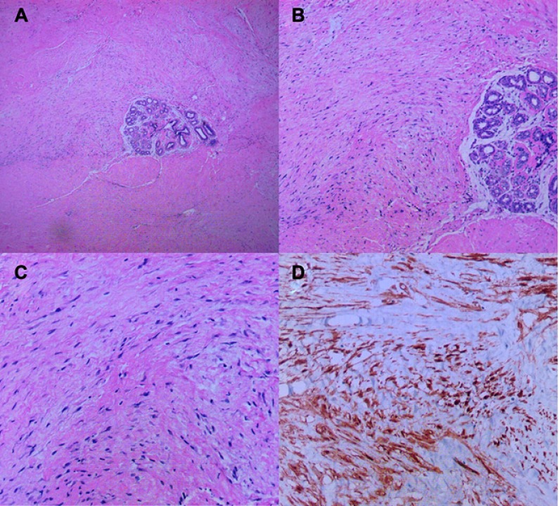
Figure 1.
H&E staining and immunohistochemical staining of specimens in the case. (A) The mass was irregular and infiltrated the adjacent adipose tissue (magnification, x40). (B) the spindle cells were arranged in parallel and partially in a wavy configuration around the mammary duct (magnification, x100). (C) The spindle cells were poorly circumscribed with eosinophilic, pale cytoplasm, sparse nucleus chromatin, and unobvious nucleoli (magnification, x200). (D) Immunohistochemical stain for β-catenin showed nuclear positivity.

