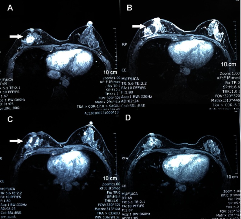
Figure 2.
Contrast enhanced magnetic resonance imaging of the breast. (A) Before treatment, a well-defined mass (arrow) was observed at the rear of right breast nipple which showed homogeneous strengthening on enhanced magnetic resonance imaging. (B) Five days after the first high intensity focused ultrasound HIFU ablation, a non-contrast enhancement region (arrow) showed up in the previous mass area. (C) Five days after the second treatment, the non-contrast enhancement region was enlarged with irregular boundary and garland-like peripheral enhancement (arrow). (D) Three months after the last treatment, the mass was well ablated and only demonstrated a patchy, heterogeneous architectural distortion region. The nipple retraction was improved.

