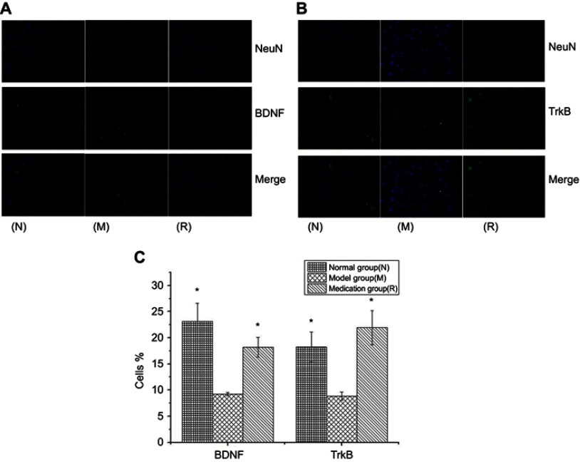
Figure 4.
DAPI staining for nuclei is in blue, and anti-BDNF (A) and anti-TrkB (B) labeled with FITC staining show neuronal cells in green; in the merged image, nuclei are in blue, and neuronal cells are in green. Histograms reporting the percentages of cells displaying BDNF and TrkB in three groups (C). N: the normal group; M: the model group; R: the medication group. Values are represented as the mean ± SEM; *P<0.05 versus M.

